Equipment
FRET-FLIM (Förster resonance energy transfer by fluorescence lifetime imaging)
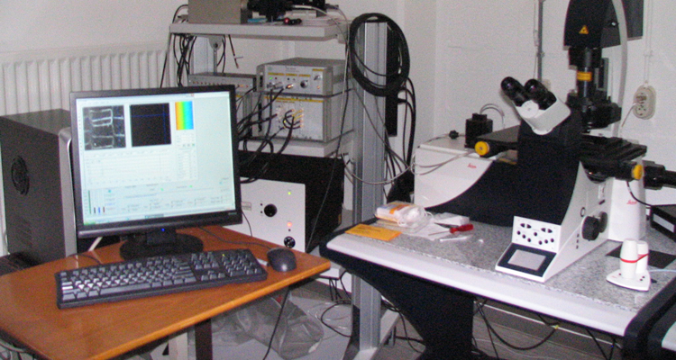
To image FRET between proteins in root cells, the Leica SP5 CLSM is equipped with fluorescence lifetime imaging (FLIM).
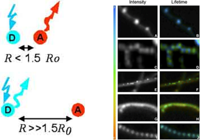
Fluorescence Resonance Energy Transfer (FRET) involves the energy transfer through dipole-dipole coupling of an donor and acceptor chromophore. The resonance conditions necessary for this process dictate that the fluorescence emission spectra of the donor overlaps with the absorption spectra of the acceptor molecule. The degree of overlap is used to calculate the spatial separation, R, for which energy transfer efficiency, E, is 50% (called the the Förster radius R0), which typically ranges from 2-7 nm. This range makes FRET an ideal mechanism for the study of protein-protein interactions and can be quantitatively determined by the measurement of fluorescence lifetime.

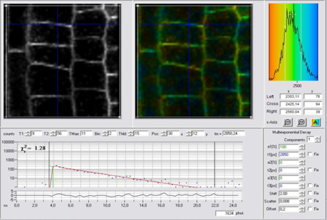
To image the occurrence of FRET between proteins in cells, the Leica SP5 CLSM is equipped with FLIM. FLIM monitors the distribution of the fluorescence lifetimes of a fluorophore at the different locations within the sample. The fluorescence of a sample is monitored as a function of time after excitation by a flash of light. We use the Time Correlated Single Photon Counting (TCSPC) method to record the fluorescence time trace for every pixel in the image. FRET is measured by the reduction of the fluorescence lifetime of the donor fluorophore.
Equipment
The Leica SP5X-SMD high-end fluorescence microscope. The instrument is based on an inverted microscope with a scanhead coupled to the base port of the microscope. It combines confocal imaging with single molecule detection (SMD). The SP5 has variable excitation with a supercontinuum tunable white light laser (WLL) source. WLL allows confocal imaging using up to eight excitation wavelengths simultaneously. Confocal microscopy with five detection channels including hybrid detector technology allows for complete, filter free, spectral freedom in imaging. Automatic 3D image acquisition using a third parameter like wavelength, time or volume.
The main feature of the SP5 is the FLIM option. Fluorescence lifetime imaging microscopy is done by the WLL tunable pulsed excitation and TCSPC detection (both Becker&Hickl and picoquant systems are available). FLIM acquisition has a time resolution of 100 ps or better and short detector dead time in combination with a high dynamic range.

Publications
- Oort, B.F. van, Marechal, A., Ruban, A.V., Bruno, R., Pascal, A.A., Ruijter, N.C.A. de, Grondelle, R. van & Amerongen, H. van (2011). Different crystal morphologies lead to slightly different conformations of light-harvesting complex II as monitores by variations of the intrinsic fluorescence lifetime. Physical Chemistry Chemical Physics, 13(27), 12614-12622.
- Krumova, S.K.B., Laptenok, S., Borst, J.W., Ughy, B., Gombos, Z., Aijani, G. & Amerongen, H. van (2010). Monitoring photosynthesis in individual cells of Synechocytis sp. PCC 6803 on a picosecond timescale. Biophysical Journal, 99(6), 2006-2015.
- Laptenok, S., Borst, J.W., Mullen, K.M., Stokkum, I.H.M. van, Visser, A.J.W.G. & Amerongen, H. van (2010). Global analysis of Förster resonance energy transfer in live cells measured by fluorescence lifetime imaging microscopy exploiting the rise time of acceptor fluorescence. Physical Chemistry Chemical Physics, 12(27), 7593-7602.
- Immink, G.H., Tonaco, I.A.N., Folter, S. de, Shchennikova, A., Dijk, A.D.J., Busscher-Lange, J., Borst, J.W. & Angenent, G.C. (2009). SEPALLATA3: the 'glue' for MADS box transcription factor complex formation. Genome Biology, 10, R24.
- Oort, B.F. van, Amunts, A., Borst, J.W., Hoek, A. van, Nelson, N., Amerongen, H. van & Croce, R. (2008). Picosecond fluorescence of intact and dissolved PSI-LHCI crystals. Biophysical Journal, 95(12), 5851-5861.
- Broess, K., Borst, J.W. & Amerongen, H. van (2007). Fluorescence lifetime imaging microscopy on individual chloroplasts in intact leaves. Photosynthesis Research, 91(2-3), 214 (PS7.17)-215.
- Almeida, R.F.M. de, Borst, J.W., Federov, A., Prieto, M. & Visser, A.J.W.G. (2007). Complexity of Lipid Domains and Rafts in Giant Unilamellar Vesicles Revealed by Combining Imaging and Microscopic and Macroscopic Time-Resolved Fluorescence. Biophysical Journal, 93, 539-553.
- Laptenok, S., Mullen, K.M., Borst, J.W., Stokkum, I.H.M. van, Apanasovich, V.V. & Visser, A.J.W.G. (2007). Fluorescence lifetime imaging microscopy (FLIM) data analysis with TIMP. The journal of statistical software, 18(8), 1-20.
- Pascal, A.A., Liu, Z., Broess, K., Oort, B.F. van, Amerongen, H. van, Wang, C., Horton, P., Robert, B., Chang, W. & Ruban, A.V. (2005). Molecular basis of photoprotection and control of photosynthetic light-harvesting. Nature, 436(7047), 134-137.