Equipment
Leica DMi8 microscope
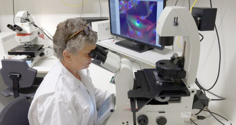
The Leica DMi8 microscope is an inverted light microscope, intended as a general microscope for routine examinations of biological specimens. This microscope will be used to make light-microscopical observations of small organisms, tissues and cells/bacteria.
The Leica DMi8 microscope can be used with normal Transparent, Fluorescence and Phase Contrast illumination. This microscope can be used with microscope slides and with culture plates, small organisms, etc. For this microscopes the LASX software is available.
The combination of the Leica DMi8 microscope with the Leica DM6b microscope and the software package in the Leica Light Microscopy Setup, makes the system well suited for both routine and research microscopy.
Technical details
Leica DMi8 microscope
The Leica DMi8 microscope is a modular inverted light microscope. The DMi8 microscope has DAPI, FITC and RHOD filter sets for fluorescence. This DMi8 has several options for transparent lighting: Bright Field, Dark Field and Phase Contrast. The Leica DMi8 is comparable with the Leica DM6b, but lacks the motorized stage.
The many automated functions allow the automatic capture of images. During the image recording process, adjustment of the viewing position (tile scanning or mark&find), and focus (extended depth of focus) are feasible. Time-lapse image capture series allow the creation of small movies of the processes of interest.
Technical functions of the Leica DMi8 as adjustment of diaphragms, condenser and the luminous intensity for the magnification and contrast method are controlled and repeated automatically. Objective-side and condenser-side IC prisms, and the analyzer and polarizer are motorized and encoded. All the motorized functions are software controlled and displayed on the touch panel Leica Smart-Touch of the DMi8. In this way the microscope can be used without the connected workstation.
Cameras
For the capturing of images three cameras are available:
- 5 megapixel and cooled colour CCD camera DFC450C,
- high-sensitive 1.4 megapixel monochrome camera DFC365FX
- fast, high-sensitive 1.3 megapixel monochrome camera DFC3000G.
The cameras can be interchanged between the two microscopes.


